Wednesday, February 20, 2008

Tissue Engineering of Oral Mocosa


Scientists at the Centre for Biomaterials and Tissue Engineering have developed an extremely valuable multi-layered epithelial model of the human oral mucosa. This is currently being used in a range of clinical treatment evaluations, as well as research into the biological mechanisms of disease.
Evaluation of the biocompatibility of dental treatments
The use of the oral mucosal model, in place of animal or patient testing, enables us to carry out very much more extensive and exhaustive test programmes. This is ideal for evaluating the biocompatibility of new dental treatments, for example, where we can test complete formulations, plus the components individually and in any combination.
The effect of any treatment can be precisely measured. Commonly we monitor cell histology, plus release of cytokines and inflammatory markers. More in-depth information can be obtained using affinity arrays to characterise gene expression in the model epithelium under control and test conditions. These tests allow us to quickly identify any irritant or toxic components on the basis of the observed changes.
Disease models
The epithelial model is being used with some success to develop an understanding of oral candidiasis ('Thrush'). We now know that an invasive, hyphal form of the fungus Candida spp. causes this. We are currently trying to evaluate what factors trigger the conversion from the relatively benign 'yeast' form that is present in most people's mouths, to the active 'disease' form.
Right: Micrographs of two sections through the oral mucosal model. The oral mucosal model at the bottom has been treated with an invasive strain of Candida spp.
Tissue engineered skin and oral mucosa are also being used to develop models of other diseases that affect these tissues. These models allow us to study disease processes and evaluate new treatments without the need for animal experiments.
Cancer models
One of the models we are developing is a model of early invasive skin and oral cancer. This project, in its early stages, is following the addition of cancerous cells to tissue engineered skin or oral mucosa. This results in an in vitro model that we hope will open a way to the development of effective treatments and better early detection of these early cancers.
Later stage tumours, and metastases, form a solid ball of cells that steadily grows in size. We can also model these in vitro by growing tumour spheroids. As the ball of cells grows in size, the center becomes starved of oxygen and nutrients. This causes tumours to release factors that encourage the growth of new blood vessels and also makes them resistant to radio- and chemo-therapy. Using tumour spheroids we can study the interaction of tumours with their surrounding tissues and the immune system as well as developing better methods for targeting treatment at these more advanced tumours.
Right: Movie clip showing the first twenty five days in the life of a breast cancer tumour. After day 6 a dark centre can be seen in the tumour ball. This is where tissue is becomming necrotic due to lack of oxygen and nutrients in the core of the tumour.
Targeted Drug Delivery
We are also using these tissue and disease models to test and develop smart drug delivery systems. By combining our knowledge of tissue specific markers and smart drug encapsulation systems we are developing the means to selectively deliver payloads of genes, radioisotopes and drugs into target cells, such as head and neck cancer cells whilst avoiding other tissues in the body. Using our model systems we can rigorously test the selectivity and effectiveness of these smart drug delivery systems before moving to clinical trials.
Researchers active in this area include:Tissue engineering of oral mucosa, use in evaluation of dental materials,

Biomaterials


There is a range of materials being developed for use in medicine, prosthetic surgery and tissue engineering. Traditionally the tough 'biomaterials' developed for prosthetics uses have been quite different from the cell culture plastics used for tissue growth 'in vitro'. Increasingly, however, surface engineering is allowing us to adjust the properties of the materials used, so that tough biomaterials and delicate culture support media can express the same surface chemistry - if desired!
Above right: A surface patterned with two different chemical groups - methyl and carboxyl - visualised by lateral force microscopy (imaging technique based on atomic force microscopy).
Working with the Sheffield Polymer Centre gives access to a wide range of new synthetic materials whose properties can be tailored to carry out specific tasks within the body or in vitro. The range of materials includes biocompatible block copolymers, dendrimers and hyper-branched polymers.
Increasingly these 'biopolymers' are being designed to be 'smart', responding to changes in their environment. Currently scientists at Sheffield can synthesise materials that change their properties in response to changes in pH, temperature, salt, ions and light. Frequently polymer structures can be tuned to undergo sharp changes in properties under conditions close to those found in the body.
Current research programmes include work to develop new materials for:
tissue engineering - as scaffolds and surfaces
controlled or targeted drug release
materials for high efficiency protein separation
DNA transfection agents
anti-fouling coatings
Many of the materials being developed are 'hydrogels', polymers that are swollen with water and can resemble living tissues in their physical properties. The water content, water structure, phase morphology, polymer structure, biofunctionality and surface science ALL affect the biological performance of these materials in natural systems - which includes cells and the proteins and other materials they secrete.

Tissue Engineering of Blood Vessel

Endothelial cells line blood vessels and regulate the passage of cells from the circulation into the tissues. This is an important 'gate keeper' function allows them to regulate many physiological and disease processes. However, this is a very dynamic process with circulating cells only interacting with endothelial under conditions of flow. Most studies of how cells interact with the endothelial lining of blood vessels are only performed under static conditions but this does not replicate what happens in life. Scientists at the Centre for Biomaterials and Tissue Engineering have therefore developed systems for studying how cells interact with the endothelial cells under conditions of flow and are using these to study a number of important disease processes.
Cancer cell binding to endothelium
Many cancers metastasise to other sites in the body. To do this, after entering the circulation, tumour cells must bind to the endothelial lining of blood vessels in order to leave the circulation and form a metastatic deposit in the tissues. This is an active process that involves the interaction of ligands on the cancer cells with adhesion molecules on the endothelium. Using flow systems we can study how different tumour cells bind to the endothelial lining of blood vessels and leave the circulation to form new cancer deposits. These studies enable us to identify markers for cancers that are more likely to metastasise and treatments that could prevent metastasis.
Right: Two looped movie clips showing fluorescently labelled cancer cells flowing over the endothelial lining of a blood vessels just as they would in the circulation. The cells in the top-most movie are from a cancer that metastasises and express a ligand that enables them to adhere to the endothelial cells. Whilst the cancer cells in the bottom movie do not express this ligand and are unable to adhere or metastasise.
Candida adhesion to endothelium
Circulating microorganisms, including bacteria and fungi, also have to bind to the endothelial lining of blood vessels in order to leave the circulation and infect the tissues. Candida albicans, is a fungal organism that is a major cause of death in patients who are immunocompromised or being treated in intensive care units. These patients are susceptible to systemic infection with this organism and once it leaves the circulation, it can cause massive tissue destruction. The mechanisms by which the organism adheres to the endothelial lining of blood vessels and invades the tissues is not known and we are using flow systems to study how Candida interacts with endothelial cells under conditions of flow.

Tissue Engineering of the Cornea


Two cornea projects are underway - addressing issues of making a biocompatible non-biodegradable replacement cornea, and of developing a carrier surface for delivering cultured corneal epithelial cells to the damaged cornea.
In the first project the challenge is to make a non-degradable, biocompatible polymeric cornea substitute, which will take the place of organ donor corneal transplants in the future. This material needs to be mechanically and optically appropriate but more exacting than this, to have a surface that promotes the attachment and migration of corneal epithelial cells. Cell attachment is a major challenge that is being tackled in a BBSRC project between Steve Rimmer in Chemistry and Sheila MacNeil in Engineering Materials. This project also involves collaboration with Dr Nigel Fullwood, University of Lancaster, who has some 10 years expertise of working on corneal epithelial cell culture.
In a second project, the University of Sheffield spin-out company, CellTran, has achieved a small business research initiative (SBRI) award from BBSRC to develop a contact lens as a carrier vehicle to deliver cultured cells from the laboratory to the patient.
Above right: Human corneal epithelial cell line cultured on (a and b) acrylic acid coated contact lenses, (c) collagen, fibronectin and BSA (positive control substrate) and (d) uncoated contact lens (negative control substrate)
Damage to the cornea can be treated by the grafting of autologous cells cultured from the contra lateral undamaged eye (if this is available) or by the use of donor corneal epithelial cells (which then requires immunosuppression). One of the challenges is how to get the cells from the laboratory to the eye and sutured onto the eye in such a way that they provide immediate cover and good take.
Currently the most commonly used method is to use human amniotic membrane. The amniotic membrane, with cells attached, is sutured onto the damaged cornea that has been denuded of cells. Clinical results with this approach are promising, but the membrane is taken from donors, and there are issues of screening and also of supply.
In the CellTran project the approach is to develop an alternative strategy, one that uses a chemically modified contact lens to transfer the corneal epithelial cells to the eye. The development of this treatment brings together two very different fields of expertise:
The development of an organ culture model (by Miss Pallavi Deshpande, 2nd Year PhD student in Professor MacNeil's group), allowing us to test the process.
Surface chemistry expertise, allowing us to treat standard contact lenses with an acrylic acid coating deposited by plasma polymerisation (by Dr Nial Bullett, Celltran), to provide a suitable transfer surface.
Bringing these together, we have recently been able to demonstrate in our model system that corneal epithelial cells can be plated onto the modified contact lens and successfully transferred from the contact lens to the eye.

Tissue Engineering of Bone


There is considerable clinical experience within the centre in the development of custom prosthetics for replacing damaged bone. Despite these modern prosthetic alternatives, however, bone remains a desirable tissue engineering target, as it has good durability and excellent biocompatibility, even when compared to modern surgical implants.
Bone regrowth within the patient's body can be achieved under favourable conditions by implanting calcium salts, possibly formed or contained within a titanium or soluble polymer mesh, directly into the patient's body. Such surgery is common in attempting to correct periodontal disease, which results in loss of bone tissue about the teeth, and eventually the teeth themselves.
Above right: A photograph of a titanium jaw implant; the mesh structure contains calcium salts to encourage bone growth through the implant.
Stem cell research is ongoing to develop therapy for inherited disorders of the skeleton and related disorders of the central nervous system. Stem cells derived from the patient can be genetically engineered to overcome the inherited disorder. The hope is that the cells can then be re-implanted into the patient, where they will assist remission of the symptoms. Studies are currently underway to see how these cells are activated and recruited in the body.
Bone is a complex tissue, which requires mechanical stimulation to develop correctly in the body. Electrospun scaffolds are being nvestigated for the culture of bone tissues in the laboratory. Experiments are underway to see how mechanical stimulation changes the nature of the tissues grown.
Mesenchymal stem cells (MSCs) are also being considered as precursors for preparing bone tissue in clinically useful quantities. It is very difficult to engineer bone directly, due to the deterioration of differentiated bone cells (osteocytes) while they are being grown in vitro. It is hoped that the MSCs, once implanted in appropriate locations in the body, will quickly differentiate to generate the required bone tissue.
An alternative technique to preparing clinically useful bone is to tissue engineer its natural precursor - hypertrophic cartilage. This cartilage is naturally mineralised in the body to produce bone. This approach has one major advantage over tissue engineering bone directly - bone requires a complex vascular system to keep the cells in it alive. Assuming a satisfactory vascular system can be developed in vitro, this must then be plumbed into the patient during the transplant to prevent areas of the bone tissue from dying within the patient. Cartilage, by contrast, normally survives without a complex vasculature, relying on diffusion of nutrients and waste products through its matrix. As a consequence fairly substantial blocks of replacement cartilage can be delivered for transplant surgery.

Tissue Engineering of Cartilage


Unlike many tissues, cartilage does not have an internal capillary network. Instead cells in this tissue acquire their nutrients and oxygen by diffusion from the surface of the tissue. As a consequence implanted cartilage generally survives well in the patient, as there is no problem with hooking it up to the patient's vascular system. This makes cartilage a useful 'halfway-house' in the development of clinically effective tissue engineering programmes - betwen two-dimensional epithelia, and tissues with complex three-dimensional organisation.
Cartilage is also a clinically important tissue, as it does not recover well from injury, and its deterioration is associated with debilitating diseases of old age, such as arthritis. Finally, cartilage is a very versatile structural tissue, different forms being used as lubricating pads in joints, as flexible support tissues in the nose and ears, and as a substrate/precursor to bone formation. The development of clinically useful cartilage through tissue engineering is therefore a useful goal in its own right.
Above right: Fluorescence micrograph showing type II collagen as a green haze about chondrocytes in engineered cartilage. This defines the extent of the chondron, the functional sub-unit of cartilage.
In nature cartilage is laid down by chondrocytes, specialised cells which are encapsulated in a chondron. The chondron, composed largely of type VI collagen, is important for protecting the chondrocyte from mechanical shock within the tissue. The complete chondron is further embedded in a matrix high in collagen type II that provides the tissue with its desirable mechanical properties.
At Sheffield scientists are developing culture techniques that give rise to a cartilage structure with composition and structure as similar to that seen in nature as possible. To do this a wide range of culture conditions have been investigated on a number of different scaffolds, including synthetic spiders' silk (which gives a good tissue).
Clinical evidence suggests that mechanical stimulation is important to the correct development of both cartilage and bone. Consequently the effect of mechanical stimulation on the growing tissue is now also being investigated at Sheffield.

Tissue Engineering of Skin


Tissue engineering of skin
Skin is an important tissue engineering target for reconstructive surgery of burns victims, but increasingly also to assist in the healing of diabetes related ulcers. The latter condition is becoming increasingly prevalent with the increased rates of late onset diabetes.
Skin is the largest tissue in the body. It is a modified epithelial tissue, with a keratinised layer of dead cells that provides a physical barrier to the outside world. Epithelial tissues have sheet-like structures, making it relatively easy to overcome problems with feeding cells during growth in vitro. While a full dermal structure, shown diagramatically below, is beyond current technology, useful engineered tissues have been developed, and are in clinical use.
Above: Diagram showing the major features of mammalian skin.
The first skin substitutes developed at Sheffield were cultured epithelial autografts. These are thin sheets of keratinocytes taken by biopsy from a patient and multiplied in the laboratory. These have been used since 1981 to treat burns victims, but they suffer a number of drawbacks:
The sheets of cells are very fragile
Take rates typically vary between 50% and 80%
They take 13 days to prepare, and half of the sheets have to be thrown away due to timing problems matching culturing the cells with clinical requirements
The sheets have to be grown on a mouse fibroblast feeder layer
Above right: A photograph of a cultured epithelial autograft; this is a delicate sheet of cells floating suspended on the growth medium in the petri dish.
In the late 90's collaboration between clinical scientists and materials scientists at Sheffield made the first big improvement on this technique - the development of flexible synthetic surfaces on which keratinocytes could be easily cultured in vitro. The synthetic support medium allows rapid culture, reducing waste, and makes the tissue very much easier to handle. The cultured keratinocytes plus the synthetic support form a flexible dressing that can be applied directly to the wound bed. Clinical studies have shown that cells migrate from the dressing to the wound and greatly accelerate healing rates, frequently resulting in complete remission for chronic ulcers that had resisted other treatments.
The technology has also been successful in treating severe burns patients. The skin dressings can be delivered within nine days, offering a valuable time saving, and large areas may be covered by contiguous application of the dressings.
Above right: An electron micrograph of keratinocytes growing on a plasma polymer surface.
A second product developed at Sheffield is a replacement oral mucosa. This is a functional replacement tissue, rather than a treatment to assist wound healing. It was developed in response to a requirement in the NHS for skin for reconstructive surgery in cases such as urethral scarring.
This tissue is based on a scaffold of sterilised skin derived from skin banks. To this are added fibroblasts and keratinocytes taken from the patient during biopsy. These cells are cultured in the scaffold, giving a tough, flexible replacement tissue. The material has been used surgically by urologists, and initial results are very encouraging.

Sunday, February 17, 2008

Function of Bone Morphogenic Protein

BMPs interact with specific receptors on the cell surface, referred to as bone morphogenetic protein receptors (BMPRs).

Signal transduction through BMPRs results in mobilization of members of the SMAD family of proteins. The signaling pathways involving BMPs, BMPRs and Smads are important in the development of the heart, central nervous system, and cartilage, as well as post-natal bone development.

They have an important role during embryonic development on the embryonic patterning and early skeletal formation. As such, disruption of BMP signaling can affect the body plan of the developing embryo. For example, BMP4 and its inhibitors noggin and chordin help regulate polarity of the embryo (i.e. back to front patterning).

Mutations in BMPs and their inhibitors (such as sclerostin) are associated with a number of human disorders which affect the skeleton.

Several BMPs are also named 'cartilage-derived morphogenetic proteins' (CDMPs), while others are referred to as 'growth differentiation factors' (GDFs).

Bone Morphogenic Protein

Bone morphogenetic protein (BMP) induces ectopic bone formation, and plays an important role in the development of the viscera. Ligand binding to its receptor induces the formation of a complex in which the Type II BMP receptor phosphorylates and activates the Type I BMP receptor. The Type I BMP receptor then propagates the signal by phosphorylating a family of signal transducers, the Smad proteins. Currently, eight Smad proteins have been cloned (Smad 1-7 and Smad 9). Upon phosphorylation by the BMP Type I receptor, Smad1 can interact with either Smad4 or Smad6. The Smad1-Smad6 complex is inactive; however, the Smad1-Smad4 complex triggers the expression of BMP responsive genes. The ratio between Smad4 and Smad6 in the cell can modulate the strength of the signal transduced by BMP.

Angiogenic Factors of Tissue Engineering

Defined as the growth of new blood vessels, angiogenesis remains an enigmatic result of cytokine action in many settings. Therapeutic stimulation of angiogenesis could relieve ischemic conditions in diseased states (e.g., peripheral vascular disease and myocardial ischemia). Currently, the following growth factors show promise for clinical applications: vascular endothelial growth factor, fibroblast growth factors, platelet-derived growth factors, transforming growth factor-β, and osteonectin. In the field of tissue engineering, induced vascularization could rescue necrotic tissue, encourage infiltration of a scaffold, and perfuse de novo tissues. Effective packaging and delivery of angiogenic growth factors, whether alone or in conjunction with biologically compatible devices, will be an important element for the successful engineering of tissue replacement technologies.

Examples of Plasmids

  1. Antibiotic resistance genes (enzymes that modify or degrade antibiotics) -- plasmids with these genes are called R factors
  2. Heavy metal resistance (enzymes that detoxify metals by redox reactions)
  3. Growth on unusual substrates (enzymes for hydrocarbon degradation, etc.)
  4. Restriction/modification enzymes (protect DNA, degrade unprotected DNA)
  5. Bacteriocins (proteins toxic to other bacteria lacking the same plasmid)
  6. Toxins (proteins toxic to other organisms; e.g. humans) -- called virulence plasmids. Some Examples:
    • Staph aureus virulence factors: coagulase, hemolysin, enterotoxin, others
    • pathogenic E. coli strains: hemolysin, enterotoxin
  7. Proteins that mediate plasmid transfer to uninfected strains

Properties of Plasmids

  • Circular DNA elements, always double-stranded DNA, Supercoiled
  • Can occur in as few as 1 copy per cell (single copy plasmids) to as many as several dozen (multicopy plasmids).


  • Variable sizes; small plasmids about 0.1% size of host chromosome, large plasmids can be as much as 10% the size of host chromosome. Smaller plasmids have few genes (30 or less). Size ranges from 1000 bp (1 kbp) to 1000 kbp.
  • Ubiquitous; almost all cells isolated in nature carry plasmids, often more than one kind. (In E. coli alone, more than 300 different plasmids isolated.)
  • View EM of plasmid DNA
  • Have a replicon (origin for DNA replication), number of copies per cell regulated. Large plasmids typically only 1-5 copies/cell (stringent control); small plasmids ~10-50 copies/cell (relaxed control)
  • Many plasmids are incompatible; if one is present, cell cannot support another plasmid of same compatibility group.
  • Not essential to cell under all circumstances; can be "cured" by agents that impair DNA replication ----> cured cell lacking plasmid. Can be spontaneously lost over time unless some selection makes plasmid valuable to cell.
  • Extend range of environments in which a cell can live (e.g., by degrading antibiotics, or providing enzymes for digestion of novel catabolites).

Generalized Transduction

  • DNA bacterial viruses = bacteriophages normally replicate in cell, produce many copies of phage DNA, many copies of phage head coat proteins, finally assemble phage heads by packing phage DNA into new phage heads. Attach tail (if present), open cell, release progeny.
    View animation of phage infection
  • Host DNA often degraded. But occasionally, piece of partially degraded bacterial DNA is correct size to be packed inside phage coat proteins, phage erroneously packs up a "mistake" = transducing phage.

  • This "mistake" phage can't cause infection; but it can be transferred to a different bacterium, get DNA into cell without risk of being degraded in environment
  • Even if this occurs, chances are slim that successful expression of DNA will occur --- still needs to undergo recombination. If most cells are killed by phage, not much use.
  • E. coli phage P1 makes this DNA packing mistake about 1 in every 1000 phage particles. Astonishingly high error rate (relative to other phages). P1 can be used as very efficient way to move small pieces of DNA from one bacterium (donor) to another (recipient). Maximum size of DNA that can fit in phage head is only about 2% of bacterial chromosome.
  • See diagram of transduction ("protected" image.) Why is this protected?

Types of Horizontal Gene Transfer

  • Natural transformation = uptake of DNA fragments from medium surrounding cell. Requires specific proteins in cell membrane, energy.
  • Only found naturally in certain cells; e.g. Pneumococcus, Hemophilus, etc. Not found in E. coli.
  • Why did this evolve? Maybe as way to scrounge DNA from dead cells, save trouble of having to make nucleotides from scratch.
  • Cells that can take up DNA are said to be competent. Requires induction of several genes. Typically occurs during exponential growth, shuts off in stationary phase.
  • Artificial transformation = laboratory technique to enhance DNA uptake of cells that do not have genetic machinery for transformation.
  • Example: treat E. coli with high Ca++ concentrations, then chill. DNA uptake now occurs (though not as well as with naturally competent bacteria).
  • Electroporation: pulsed electric fields produce short-lived membrane pores, allows DNA movement (either in or out of cell). Useful way to move small pieces of DNA and plasmids between cells.
  • See natural transformation and artificial transformation ("protected" image.)

Horizontal Gene Transfer

Horizontal gene transfer (HGT), also Lateral gene transfer (LGT), is any process in which an organism transfers genetic material to another cell that is not its offspring. By contrast, vertical transfer occurs when an organism receives genetic material from its ancestor, e.g. its parent or a species from which it evolved. Most thinking in genetics has focused on the more prevalent vertical transfer, but there is a recent awareness that horizontal gene transfer is a significant phenomenon.

There is some evidence that even higher plants and animals have been affected. Dr. Mae-Wan Ho, a noted scientist and critic of genetic engineering, writes: "While horizontal gene transfer is well-known among bacteria, it is only within the past 10 years that its occurrence has become recognized among higher plants and animals. The scope for horizontal gene transfer is essentially the entire biosphere, with bacteria and viruses serving both as intermediaries for gene trafficking and as reservoirs for gene multiplication and recombination (the process of making new combinations of genetic material)." But Richardson and Palmer (2007) are more cautious: "Horizontal gene transfer (HGT) has played a major role in bacterial evolution and is fairly common in certain unicellular eukaryotes. However, the prevalence and importance of HGT in the evolution of multicellular eukaryotes remain unclear.

Due to the increasing amount of evidence suggesting the importance of these phenomena for evolution (see below), molecular biologists such as Peter Gogarten have described horizontal gene transfer as a "A New Paradigm for Biology".

It should also be noted that the process is emphasised by Dr. Mae-Wan Ho as an important factor in "The Hidden Hazards of Genetic Engineering", as it may allow dangerous transgenic DNA (which is optimised for transfer) to spread from species to species.

The Bioartificial Pancreas

Instead of using genetic therapy, as Dr. Block suggests, foreign cells that secreted a particular protein could be used to replace the patients own-- if a way was found to protect the cells from the host's immune system. This is exactly the idea behind the "bioartificial pancreas" which researchers hope will replace insulin injections as a therapy for diabetics. The insulin-secreting cells form pancreatic islets, usually taken from a pig, are placed in semi-porous capsules and implanted in the body. The capsules must be biologically and chemically inert; that is, their chemical composition cannot induce inflammation or other reaction from the body, and they must resist decomposition. The capsules must contain pores small enough to exclude the mobile cells of the immune system, macrophages and lymphocytes, but large enough to allow a physiological release of insulin in response to blood glucose levels. Obviously, long term survival of the cells is also a requirement. According to Anthony Sun, of the University of Toronto, diabetic monkeys have become insulin independent using the bioartificial pancreas for periods longer than two years. Other researchers have shown similar results in dogs and rats. Islet cells have been recovered after a year of implantation and shown to be still viable. A limited trial using human beings that was carried out in China was so successful, according to Dr. Sun, that patients tried to bribe the doctors to continue the experimental procedure. Clinical trials have now been only recently approved by the FDA in the United States using microencapsulated porcine cells in human patients.

Genetic Therapy

Dr. Block is excited about his experiments for another reason. The liver has long been thought of as an ideal target for genetic therapy; it is the largest organ in the body, , and it is specialized for delivering secretion products into the blood stream. Suppose, for instance, that a patient is hemophiliac, that is, he doesn't produce blood clotting factor, a protein that is encoded by a gene, a piece of DNA. A portion of his liver could be removed, a procedure which would do him no harm, as the liver would regenerate to full size (virtually the only organ with that capability). The excised liver cells could then be genetically engineered such that a new gene specifying the clotting factor protein was introduced. The cells could then be re-implanted in the liver, and clotting factor would then be secreted by engineered cells. The problem, so far, has been that hepatocytes would not undergo cell division outside the body. Cells that are not actively replicating their chromosomes will not take up and express the genetically engineered DNA used by molecular biologists for genetic therapy. If Dr. Block has persuaded liver cells to divide in culture, this problem may be solved.


The Bioartificial Liver

There is reason for optimism that the bioartificial liver can eliminate the need for a transplanted liver in other patients with fulminant liver failure. Ironically, nobody knows what toxins accumulate in the brain, or how the liver operates to relieve intracranial pressure. "We are treating a disease we do not understand with a treatment we do not understand," according to Demetriou. Acute liver failure is usually due to a temporary event, like Tylenol overdose (a common means of attempted suicide) or hypersensitivity to other drugs. The liver is a fairly robust organ and could possibly regain function in many instances--if the patient doesn't go brain-dead first. This is an important point, one he hopes to impress on health maintenance organizations (HMOs) and insurance companies. Traditionally, these companies have been slow to adopt payment schedules for new technologies. Demetriou's bioartificial liver treatments require constant monitoring in expensive intensive care units. Worse yet, from the financial side, the treatments allow a patient who might have died access to a liver transplant, which costs in excess of half a million dollars. If Demetriou can show recovery of patients, in some cases, without a liver transplant, he stands a far better chance of having the bioartificial liver accepted by HMOs, Medicare and private insurance companies as standard medical practice in the U.S. For every 7 hours of treatment with the bioartificial liver, Demetriou figures he gains another 24 hours of life for his patients. Although the experiment hasn't really been tried yet, most in the field expect that using pig liver cells instead of human limits the amount of time that the device could be used on a human patient. In addition to its detoxification function, the liver supplies most of the non-cellular protein found in the blood. It is expected that eventually the patient will develop an immune response to the pig proteins secreted by the pig liver cells-- and Demetriou concedes he has detected porcine proteins in the blood of his patients.

Long term treatment of patients with chronic liver disease in a manner analogous to the hemodialysis treatment of kidney patients would probably require a bioartificial liver which used human cells. But where to get the cells? As it stands now, technology is not the limiting factor in treating liver patients; it is the number of human livers available. Every year, 30-50,000 people die of liver failure, while only about 3000 transplants are available. Geoffrey Block may have an answer to the human liver shortage. Recently, he and his co-workers at the University of Pittsburgh stimulated liver cells to divide and grow in laboratory culture, using a chemically defined culture medium. The secret? A few hormones--some of which are proprietary, and 75 different nutrients. Surprisingly, fully differentiated, functioning hepatocytes divide and grow, according to Block. It had been widely believed by many cell biologists and embryologists that these fully differentiated cells will not reproduce. Differentiated cells are replaced, it is thought, by the division of immature "committed progenitor" cells. While the latter can be cultured in the laboratory, they lack the liver cell functions that would be needed in a bioartificial liver. Block believes that his group has "dedifferentiated" the liver into growing, immature cells. He also claims that he can manipulate the cultures to develop again into functioning mature hepatocytes, which would be necessary for his work to be medically significant. Lola Reid of the University of North Carolina, who has done careful work on the developmental biology of liver, urges caution in interpreting Block's results. The "de-differentiation" of hepatocytes runs contrary to her own experience, which suggests that the liver cells mature in an ordered fashion in the body, starting with the division of immature stem cells. Demetriou, for his part, is skeptical that Block's laboratory cultured cells will ultimately provide the detoxification function necessary for use in the bioartificial liver, citing many previous claims in the literature of laboratory cultured, functioning hepatocytes that turned out to be premature. If Block is correct, however, "I'll buy the cells from him," says Demetriou.

From Tissue to Organs

Successes in regenerating skin and cartilage have led scientists to imagine the possibility of regenerating or reconstructing vital organs, for example, the liver. The problems are more difficult because the three dimensional architecture is complex and contains an assortment of different tissue types. The constituent cells will not automatically assume the correct structure without a matrix to guide them. Griffith-Cima has turned to a technique called "three-dimensional printing" (3DP) to create a biodegradable scaffold, which she is using to recreate miniature livers in the laboratory. 3DP is a computer aided design, computer aided manufacturing (CAD/CAM) technology wherein a three-dimensional blue-print programmed or scanned into the computer is translated into a three-dimensional structure by alternating layers of powder and adhesive in a precisely controlled fashion. Although Griffith-Cima admits it sounds like a Star-Trek replicator, it is actually more akin to the common ink-jet printers found in many offices, only engineered to work in three dimensions instead of two. Originally developed for ceramics, Griffith-Cima has altered 3DP to work with biodegradable suture-like materials (e.g. polyglycolic acid).

Griffith-Cima's "mini-liver" scaffolding has an "artery" at one end and a "vein" at the other, with a branching network in-between. So far, she has been able to show that liver cells seeded on to her scaffold will sort into "hepatocytes" , cells with the metabolic functions of the liver, and "endothelial cells", which line the capillaries and blood vessels, in a manner similar to what would be expected for a natural liver. Eventually, she hopes to have a laboratory generated liver which can be implanted in patients with liver dysfunction, reducing the need for liver transplants from organ donors. Occasionally, for inspiration, Griffith-Cima tours the wards of Childrens Hospital with her collaborator, Joseph Vacanti. She will regard her work as successful, she says, the first time a child is saved when one of her experimental livers. Vacanti is convinced that the laboratory cultured liver "is the best long term solution."


Tissue in Three Dimension

As important as it is, skin is basically a two-dimensional tissue, whereas most of the tissues and organs in our body are more complicated. Can engineers make tissue in three dimensions? Last year, as a test of this proposition, Linda Griffith-Cima and her colleagues at MIT created tissue that looked very like a human ear The rationale for this project was provided by Griffith-Cima's collaborator, Dr. Joseph Upton, a plastic surgeon who is frequently in the position of trying to reconstruct an ear for his patients. The outer ear is composed of cartilage and skin. Cartilage is synthesized by a specialized cell called a chondrocyte. Essentially, cartilage is extensive extracellular matrix material secreted by chondrocytes, and composed mainly of collagen, but also including large, spongy carbohydrate chains (glycose amino glycans) as well as some minor proteins. Griffith-Cima seeded isolated chondrocytes onto an artificial matrix composed of biodegradable suture material, shaped into the form of a human ear. After the chondrocytes attached to the matrix in the laboratory, the whole construct was transplanted under the skin on the back of a mutant laboratory mouse, called a nude mouse. In addition to having no hair, nude mice are lacking an immune system organ called the thymus, and as a result, they are almost totally immunodeficient. Nude mice will accept almost any type of graft (researchers have grown feathers on nude mice!). Once implanted in the mouse, the chondrocytes produced cartilage, dissolving the synthetic matrix in the process. The result: an apparent human ear growing out of the back of the mouse! It quickly became "the ear seen round the world."

For the aggressive leader of ATS, Gail Naughton, the press attention given to the vivid image of the ear seems to represent a challenge, or perhaps an opportunity to promote her company, which is also pursuing the possibilities of laboratory engineered cartilage. At the recent BioArtificial Organs conference, Naughton presented her response to the ear on the mouse's back--a very human looking ear implanted into a rabbit where the rabbit's floppy ear ought to be. The effect is grotesque, and calls up sci-fi warnings of technology gone awry. But the message gets through. Yes, tissue in three dimensions can be constructed, and yes, it is stable when placed in the animal (and presumably in a human being). The "human" ear on the rabbit was actually constructed from rabbit chondrocytes--human chondrocytes would have been rejected by the rabbit. The human ear shape was supplied by the synthetic matrix on which the cells were originally grown, and is retained in the transplanted animal even though the original biodegradable matrix has been dissolved and replaced with cartilage. Thus, "rabbit ear shape" is not an inherent property of rabbit chondrocytes. Shape is a property that can be "taught" by the matrix on which the cartilage secreting cells are grown.


Regenerating Skin

Skin is an important vital organ, a very effective, breathable, moisture seal, without which our wet, pulpy insides would immediately dry out. Skin also protects us against all the bacteria, mold, and parasites for whom the human body represents opportunity. The injuries of severe burn patients must quickly be covered and traditionally, this has involved the use of cadaver skin. Obviously, this treatment brings with it the risk of viral infection and is very expensive and time-consuming; sometimes the cadaver skin has too be stitched together from small segments. John Burke was looking for a way to avoid this ghastly business for his patients and himself. He hoped to create an artificial covering that would take the place of cadaver skins. His first attempts to do this were, in his words, "the most glorious messes...absolute catastrophes." Burke needed someone who understood materials. Ioannis Yannas wanted to associate with "a medical person, who would be in contact with the patient population, who would know the exact needs of that population, so I could design a product with those specifications in mind." The surgeon and the chemist , together, invented the "skin regeneration template" which Integra now markets as "Artificial Skin." Artificial Skin far exceeds John Burke's initial expectations. Instead of merely being a temporary covering protecting the patient, Artificial Skin actually allows the body to create a new dermis over the burn site.

Artificial Skin is made in two layers; the upper layer is an elastic silicon membrane which provides the moisture barrier, functionally replacing the epidermis, and the lower level consists of an synthetic matrix of collagen fibers (purified from bovine tendons) and chondroitin sulfate (a type of large carbohydrate extracted from shark cartilage). This synthetic matrix was designed to approximate the supporting layer of protein and carbohydrate normally secreted by dermal cells. Dermal cells, as well as most tissues in the body, exist spread on surface called an "extracellular matrix" consisting of proteins and carbohydrates. Collagen is the major protein found in extracellular matrices in the body. It is also the protein from which we make gelatin. Because the protein is in a disordered state in gelatin, it has no particular structural strength. In extracellular matrices, however, collagen exists as a long, stretched out protein that arranges itself in triple helical bundles, similar to rope or twisted cable, although on a microscopic scale. Along these bundles are numerous small attachment sites for cells. Unexpectedly, Burke and Yannas found that when the synthetic matrix supplied by Artificial Skin was applied to a wound site, dermal cells from around the wound site migrate into to the artificial matrix and attach to the collagen fibers. The bovine collagen is slowly degraded and replaced with authentic human collagen synthesized by the dermal cells. Blood vessels grow into the wound to vascularize the new tissue. After the dermal layer has had a chance to repair itself, the outer membrane can be removed and replaced with very thin epidermal transplant, once again providing a natural moisture seal. The resultant skin is functionally and cosmetically superior to that achieved with other methods of treatment.

Is there any magic formula for making a collagen matrix that will attract dermal cells to colonize it? "Yes, and it is magical in a way that doesn't make sense to most biologists I know," says Dr. Yannas, the chemist. The collagen must have a "specific surface" which involves the right density of attachment sites for cells. The rate at which the matrix degrades is also very important. The matrix must persist while the "inflammation rages on," but ultimately it must be degradable so that it can be replaced by authentic human collagen. Additionally, the bovine collagen must be treated to remove or mask immunogenic sites that might cause the body to reject Artificial Skin in the same way that it would reject a foreign skin graft, for instance.

Artificial skin illustrates some of the general principles involved in tissue engineering. Tissue is organized by the underlying extracellular matrix. The matrix itself, was originally secreted by the cells, themselves, or put in place by their predecessors during the process of embryogenesis. In a deep wound, such as a severe burn, the protein matrix itself is missing or severely damaged, and the original cells in the wound have died. The burn site itself is temporarily occupied with immune system cells, like macrophages and lymphocytes, which keep infection from spreading but have no innate ability to manufacture skin. There are no appropriate cells left to replace the matrix correctly, and there is no matrix left to organize the tissue. Artificial Skin solves the problem by providing a synthetic matrix, or scaffolding, on which new tissue can arrange itself. Integra claims to have had excellent results at healing burns with Artificial Skin, even in older patients, whose skin is already thin and brittle with age. So pleased are they, in fact, according to vice-president Robert Towarnicki, that Integra is now conducting clinical trials to expand its use to cosmetic plastic surgery, that is, to treat scarring caused by previous wounds or burns. This is, of course, a vastly larger market than the original indication.

Although Integra is the first to gain FDA approval, two other biotechnology companies, Advanced Tissue Sciences (ATS) and Organogenesis, are also advancing their own form of engineered skin. Both of these companies actually grow living human "skin" in the laboratory, and use it to patch the sites of wounds. The ultimate source of their skin cells is human infantile foreskins harvested by circumcision. The cells in foreskin will grow and divide in tissue culture, increasing in number, if given an appropriate medium containing nutrients and growth factors. Infant foreskin has more potential for cell division than does that from an adult; cell cycle time increases with age, and the ultimate number of divisions is finite. An infant's foreskin can theoretically grow into a lawn that would cover something on the order of six football fields! (assuming you could incubate football fields in a humidified, sterile chamber at body temperature in a pH buffered solution containing vitamins, amino acids, epidermal growth factors, insulin, glucose, etc.) ATS has not had to acquire a new foreskin since 1989, despite ongoing clinical trials of its products. It's Dermagraft-TC would compete with Integra's Artificial Skin for the same indications, given FDA approval.

ATS and Organogenesis are also addressing the acute need for treatment of diabetic ulcers. Ulcers in the extremities, particularly the feet, result from poor circulation, very common in diabetic patients. ATS estimates that 500,000 patients are treated per year, and that over 55,000 amputations are performed because of inability to heal the wounds. ATS is allied with Smith and Nephew, PLC, a British pharmaceutical giant with over $1.5 billion in sales. The deal with Smith and Nephew is illustrative of the hurdles that small biotechnology companies face. ATS got $10 million up front and will receive $5 million more assuming they win FDA approval of Dermagraft for diabetic ulcer treatment. FDA pre-market approval of a medical device, however, does not obligate Medicare or private insurance companies to pay for that product, an important point in this age of dwindling budgets and managed health care. ATS will receive another $5 million if they successfully lobby Medicare into approving reimbursement for their product. Additional funds, up to $40 million would be given, pro-rated according to gross sales achieved, according to Marie Burke, director of investor relations for ATS.


Engineering Human Tissue

If a railroad engineer runs a freight train, an electrical engineer builds electric circuits and a custodial engineer is --well, a janitor-- what then is a tissue engineer? Engineer is one of those English words that have gotten slippery in their application. The Middle English engin comes from the Latin root ingenium for natural ability, or genius. Natural abilities were somehow ascribed to machines, called engines, the operators or designers of which were called engineers. This confusion between the natural and the mechanical is getting worse. Tissue engineers, the subject of this article, are people that design and build bioartificial organs among other things. Bioartificial, a word with oxymoronic qualities, refers to synthetic organs that contain living cells or tissues. A bioartificial liver, for instance, is a device which contains liver cells to detoxify the blood of a patient, along with purely mechanical filters, tubes and pumps.

Tissue engineering, broadly defined, covers a large fraction of the problems that medical science encounters. It includes 1) inducing the patient's own body to regenerate damaged tissue; 2) replacing the patients' cells or organs with living tissue from other sources, or 3) implanting prosthetic devices, such as an artificial heart, which functionally replace living organs or tissues. Already, physicians have found ways to enhance the body's limited abilities in the regeneration of skin, bones, cartilage, and nerves. Bioartificial livers, pancreas, and kidneys are in various stages of clinical development. Robert Langer and Joseph Vacanti, among the visionary fathers of tissue engineering, imagine a future in which severed limbs could be fully regenerated, failed organs could be replaced with living, laboratory generated equivalents, blindness could be cured with microelectronic vision chips, and fetuses could be brought to term in artificial wombs. This is not a far out future but one that could be realized in 30-50 years, in their view, given recent advances in understanding of tissue and organ culture, and in synthesizing biocompatible materials.

Technology, like politics, is the art of the possible. Advances in science are a necessary but not sufficient condition for advancements in technology. Also necessary are commitments of time and money, cultural adjustments in attitude, and sometimes, enabling changes in the regulatory environment.

The delicate balancing act required for technological advance is demonstrated by a small biotechnology concern called Osteotech in its efforts to help bone surgeons. While bones have an inherent ability to repair themselves, particularly traumatic fractures or bone cancer can leave voids that are impossible to for the body to heal in a satisfactory fashion. Traditionally, these voids have been filled with grafts, usually from taken from the patient; for instance, a piece of the hip is often used in back surgery. This requires the pain and expense of a second operation, however, and in sometimes the patient is not an adequate self-donor. In that case, "allograft" tissue, (in this case, bone) from another human donor is used, usually from a cadaver. Osteotech slices, freezes, and packages human bone in sizes convenient for surgeons.

The main attribute of bone, its structural strength, also makes it a hard material to work with. For this reason, Osteotech supplies Grafton, which comes in flexible strips, as a gel, or even Grafton bone putty. Grafton is "demineralized bone matrix;" human bone that has been chopped up, freeze-dried, acid extracted and further processed into its various forms. Grafton, however, is not a product that Osteotech "sells" to end users, because, under U.S. law, human tissue cannot be bought or sold. Technically speaking, Osteotech "processes" human tissue for non-profit organizations, such as the Red Cross or the Musculoskeletal Foundation, which then distribute it to surgeons. Last year, the federal Food and Drug Administration introduced an additional complication when it considered at length whether Grafton should be regulated as a "medical device" , and hence under FDA control. To the considerable relief of Osteotech and its stockholders, the FDA eventually concluded that Grafton was a human tissue graft ( regulated by local institutional review boards (IRBs)), thus saving the years and expense that would have been required to obtain FDA approval. It is likely that Grafton might easily be replaced with bone matrix from non-human sources, such as cattle, at much lower expense. Ironically, such a product, by the same logic that the FDA applied to Grafton, would be considered a "xenograft" or a tissue donation of non-human origin, which the FDA now regulates in a recent extension of its powers. Therefore, we are unlikely to see such a non-human derived Grafton-like product marketed in the U.S. very soon, if ever.

cal devices" whi

Natural Breast Implant


The implant methods used by modern breast reconstructive and cosmetic surgery rely on artificial implants consisting of bags of silicone or saline solution. Silicone/saline solution implants, however, have not presented a strong safety record, since there were many cases of rejection or rupture of the implant. Not to mention that they tend to lose their shape after a few years, thus not being suitable for lifetime use.
This may change drastically within the next decade, as scientists at the University of Illinois, Chicago presented, at the American Association for the Advancement of Science conference in February, successful results of recent research concerning natural implants, made of tissue artificially grown from human stem cells.

Stem cells are undifferentiated “master cells” that have the ability to grow into any one of the body’s more than 200 cell types, under the appropriate conditions. By mimicking the exact natural conditions in which fat cells develop in the human body, the research team successfully created and then multiplied adipose tissue cells in the laboratory, placing them onto special hydrogel scaffolding, in order to properly nourish them and mold them into shape. This process completed, the implant material created is ready to be inserted into the body.

Since the material inserted is created from the patient’s own stem cells, it will constitute no risk of rejection, unlike artificial implants made of silicone or saline solution. Vascularization of the implanted material will also become possible. The researchers believe that this technique has the potential to revolutionize other reconstructive types of surgery as well, other from breast surgery, particularly due to the fact that the implant material is capable of maintaining its size and shape for a lifetime, as compared to conventional implants, that lose between 40% and 60% of their volume over time. Last but not least, there is obviously no risk of rupture or leakage.

Even though the technique has only been tested on mice, once the viability of grown implants is proved for humans as well, the implants grown from stem cell material could become largely available within a decade.

Next page >>>

What is Polyhydroxyakanoates for Tissue Engineering?

As PHA composes of various hydroxyalkanoates that are synthesized by as many as 75 different genera of gram-positive and gram-negative bacteria, an extensive group of properties are available (Reddy et al 2003). Non-storage PHA, poly(3HB) have been found in the cytoplasmic membrane and cytoplasm of Escherichia coli, as well as in yeasts, plants and animals. The mechanical and biocompatibility of PHA can also be changed by blending, modifying the surface or combining PHA with other polymers, enzymes and inorganic materials, making it possible for a wider range of applications There are several different pathways for PHA formation

Windpiped Defect Repair Possible

It's been across the world's press, yes, the report by Dr. Fauza's group in the Boston Children's Hospital.

The team have employed a method of seeding embryonic cells onto a biodegradable scaffold, and with the addition of growth factors, the cells produced healthy cartilage. The grafts, once mature, were used to repair the windpipes of seven foetal lambs, animals which grow at approximately the same rapid rate as a human.The technology, termed autologous chondrocyte implantation, has been previously employed within the cartilage of joints, with mixed success. The findings of this study are almost a best case scenario - the cells are young and strong from the amniotic fluid, and the lamb's body at a foetal stage is surging with growth hormones, proteins, and a very particular healing process able to work much more effectively than that in an adult.

The age of such cells is very important, every time cells divide, they become less metabolically active, and more likely to become cancerous. Most cells exhibit senescence, a property by which they will eventually stop dividing. Rapid growth of cells from an adult in order to perform this kind of proceedure may result in an increased risk of cancer, or at the best, impaired future tissue health.

The healing within both a foetus and a neonate is very rapid, and rarely leaves a scar. The process itself is so different, that doubt may be raised as to whether the healing response observed in this study could be replicated in other than paediatric cases.

All in all, these are excellent results for a particular problem, and highlight the vast body of research currently taking place on this elegant combination of natural healing and high technology.

Tissue Engineered Heart Repair

There are a wide range of treatments in active use for such cartilage defects, although many of them have questionable or untested benefits which have not seen the light of rigorous scientific investigation. Within this category fall lavage and debridement. Microfracture and abrasion have been shown to produce some form of repair, but it itself is somewhat short-lived


The current research emphasis is very-much aimed at the use of Tissue Engineering for cartilage. Tissue Engineering is a strategy which combines living cells, bio-materials, and an appropriate range of mechanical and/or biological stimulation to replace or improve tissue functionality with a living biological repair.Using mice models and sophisticated gene chip technology, they found that mice that lacked certain specialized proteins had worse heart function and a decreased heart repair capacity after heart attacks as compared to mice that had these proteins, called stem cell factor receptors.

These stem cell factor receptors are frequently found on stem cells in the bone marrow and their role is to activate other "signaling" molecules, which cause stem cells to leave the bone marrow and migrate via the blood stream to the damaged heart tissue.

A further proof that stem cell factor receptor signaling works this way is the fact that once the knock-out mice underwent bone marrow transplantation from normal mouse donors, cardiac function was improved.

What is Cartilage Tissue Engineering?

The human articulating joints, particularly those of the lower limbs, are able to last a lifetime, with good care, and good luck. All-too-often, however, either through traumatic injury or a simple over-use, the cartilage surface of the joint starts a slow and steady decay. This decay, often described as abnormal turnover in which the destruction of cartilage exceeds the production, eventually leads to painful joints and a loss of mobility.
There are a wide range of treatments in active use for such cartilage defects, although many of them have questionable or untested benefits which have not seen the light of rigorous scientific investigation. Within this category fall lavage and debridement. Microfracture and abrasion have been shown to produce some form of repair, but it itself is somewhat short-lived.


The current research emphasis is very-much aimed at the use of Tissue Engineering for cartilage. Tissue Engineering is a strategy which combines living cells, bio-materials, and an appropriate range of mechanical and/or biological stimulation to replace or improve tissue functionality with a living biological repair.

Advantages and Disadvantages of Tissue Engineering

There are several treatment options for organ failure or tissue loss – transplants, reconstructive surgery, artificial prosthesis or mechanical devices (kidney dialyzers, prosthetic hip joints, mechanical heart valves), but unfortunately, they are imperfect. There's a declining availability of organs, there's the need for multiple surgery in the case of autografts and mechanical devices do not have the capacity to perform all functions of an organ. Prosthetic replacements present risks such as thrombosis, an increased susceptibility to infection, limited durability, need for reoperations.In this context, the emergence of the science called tissue engineering is more than just salutary. The purpose of tissue engineering is to create tissues in culture for use as replacement tissues for damaged body parts. Within the past 10 years, the creation of bioartificial tissues has achieved a series of successes.

The science of tissue engineering combines the principles of bioengineering, cell transplantation, hematology ad those of material science/engineering, for the unique goal of generating bioartficial tissues and organs. Skin, cartilage and bone have been synthesized in the laboratory, and success has been predicted in the creation of blood vessels, blood and organs such as heart, lungs, pancreas, and liver. Attempts have been made to create artificial corneas, intestines and heart valves. Bladders have been bioengineered and implanted in dogs, with total success.

The process generally comprises of the isolation of cells from a patient and their growth on three-dimensional templates or scaffolds (matrices), under the conditions necessary for them to develop into functional tissue. Then, the tissue-biomaterial construct is implanted into the patient. The biomaterial gradually absorbs, ensuring that only the natural tissue remains in the body, having acquired the shape of the material. This process completed, the bioartificial tissue becomes structurally and functionally integrated into the body.

What is Pluripotent Cells?

Pluripotent Cells


In humans,‭ ‬after the first four days,‭ ‬the cells are no longer able to produce every type of tissue required‭; ‬they have already begun to differentiate.‭ ‬At this stage they can already be broadly grouped into the three primary germ layers‭; ‬the‭ ‬ectoderm‭ (‬covering tissues such as skin and retina‭)‬,‭ ‬endoderm‭ (‬the gastrointestinal tract,‭ ‬respiratory tract,‭ ‬the liver and the pancreas‭) ‬and the‭ ‬mesoderm‭ (‬mostly connective and circulatory,‭ ‬such as bones,‭ ‬heart,‭ ‬blood vessels and muscles‭)‬.‭ ‬As these tissues are able to produce multiple cell-types but are not able to produce all cell-types they are termed pluripotent,‭ ‬from the latin‭ ‬pluralis meaning more.

Stem Cells


The totipotent and pluripotent cells where sourced from an embryo are a classified as‭ ‬embryonic stem cells,‭ ‬cells within the embryo from which other types of cell originate.

What is Totipotent Cells?

Totipotent Cells


From the zygote which divides to form an embryo springs forth every type of cell in the body,‭ ‬of which there are over‭ ‬200,‭ ‬and all the tissues required for the placenta.‭ ‬Because of this,‭ ‬these very early cells which,‭ ‬in humans,‭ ‬occur just in the first four days following fertilisation,‭ ‬are termed totipotent.‭ ‬Toti-‭ ‬is from the latin‭ ‬totus,‭ ‬meaning whole‭ (‬the same root of the word total‭)‬,‭ ‬and potent being from the latin‭ ‬potens meaning to be able‭; ‬thus the cell is able to produce any other cell.‭

Treatment of Tissue deficiency

The treatment of tissue deficiency is currently constrained,‭ ‬if not by financial implications,‭ ‬by the limited range of approaches to treatment currently available in most cases.‭ ‬Once serious tissue damage has taken place it is unlikely that spontaneous recovery will take place other than in a few isolated cases.‭ ‬Mechanical solutions can help in some cases,‭ ‬such as a heart-lung machine or a crutch,‭ ‬but these rarely restore functionality in a satisfactory and long-lasting manner.‭ ‬This means that transplantation has become the mainstay of treatment,‭ ‬a strategy which has always been and is always likely to be significantly restricted by a lack of donor organs and tissues and dangers associated with tissue rejection and the transmission of disease.

Tissue Engineering thus aims to provide an alternative better means of treatment for tissue and organ damage through combining both biological and artificial components in such a way that a long-lasting repair is produced.‭ ‬This rapidly-emerging field is strongly interdisciplinary as it combines engineering techniques,‭ ‬materials science and biochemical expertise.

Bioreactors

In many cases, bioreactors are employed to maintain specific culture conditions. The devices are diverse, with many purpose-built for specific applications. Bioreactors allow for precise and continuous control of culture conditions and also allow for introduction of different stimuli to tissue cultures.


Assembly Method

One of the continuing, persistent problems with tissue engineering is mass transport limitations. Engineered tissues generally lack an initial blood supply, thus making it difficult for any implanted cells to obtain sufficient oxygen and nutrients to survive, and/or function properly.

Self-assembly may play an important role here, both from the perspective of encapsulating cells and proteins, as well as creating scaffolds on the right physical scale for engineered tissue constructs and cellular ingrowth.

It might be possible to print organs, or possibly entire organisms. A recent innovative method of construction uses an ink-jet mechanism to print precise layers of cells in a matrix of thermoreversable gel. Endothelial cells, the cells that line blood vessels, have been printed in a set of stacked rings. When incubated, these fused into a tube.

Synthesis of Tissue Engineering scaffold

A number of different methods has been described in literature for preparing porous structures to be employed as tissue engineering scaffolds. Each of these techniques presents its own advantages, but none is devoid of drawbacks.

  • Nanofiber Self-Assembly: Molecular self-assembly is one of the few methods to create biomaterials with properties similar in scale and chemistry to that of the natural in vivo extracellular matrix (ECM). Moreover, these hydrogel scaffolds have shown superior in vivo toxicology and biocompatibility compared with traditional macroscaffolds and animal-derived materials.
  • Textile technologies: these techniques include all the approaches that have been successfully employed for the preparation of non-woven meshes of different polymers. In particular non-woven polyglycolide structures have been tested for tissue engineering applications: such fibrous structures have been found useful to grow different types of cells. The principal drawbacks are related to the difficulties of obtaining high porosity and regular pore size.
  • Solvent Casting & Particulate Leaching (SCPL): this approach allows the preparation of porous structures with regular porosity, but with a limited thickness. First the polymer is dissolved into a suitable organic solvent (e.g. polylactic acid could be dissolved into dichloromethane), then the solution is cast into a mold filled with porogen particles. Such porogen can be an inorganic salt like sodium chloride, crystals of saccharose, gelatin spheres or paraffin spheres. The size of the porogen particles will affect the size of the scaffold pores, while the polymer to porogen ratio is directly correlated to the amount of porosity of the final structure. After the polymer solution has been cast the solvent is allowed to fully evaporate, then the composite structure in the mold is immersed in a bath of a liquid suitable for dissolving the porogen: water in case of sodium chloride, saccharose and gelatin or an aliphatic solvent like hexane for paraffin. Once the porogen has been fully dissolved a porous structure is obtained. Other than the small thickness range that can be obtained, another drawback of SCPL lies in its use of organic solvents which must be fully removed to avoid any possible damage to the cells seeded on the scaffold.
  • Gas Foaming: to overcome the necessity to use organic solvents and solid porogens a technique using gas as a porogen has been developed. First disc shaped structures made of the desired polymer are prepared by means of compression molding using a heated mold. The discs are then placed in a chamber where are exposed to high pressure CO2 for several days. The pressure inside the chamber is gradually restored to atmospheric levels. During this procedure the pores are formed by the carbon dioxide molecules that abandon the polymer, resulting in a sponge like structure. The main problems related to such a technique are caused by the excessive heat used during compression molding (which prohibits the incorporation of any temperature labile material into the polymer matrix) and by the fact that the pores do not form an interconnected structure.
  • Emulsification/Freeze-drying: this technique does not require the use of a solid porogen like SCPL. First a synthetic polymer is dissolved into a suitable solvent (e.g. polylactic acid in dichloromethane) then water is added to the polymeric solution and the two liquids are mixed in order to obtain an emulsion. Before the two phases can separate, the emulsion is cast into a mold and quickly frozen by means of immersion into liquid nitrogen. The frozen emulsion is subsequently freeze-dried to remove the dispersed water and the solvent, thus leaving a solidified, porous polymeric structure. While emulsification and freeze-drying allows a faster preparation if compared to SCPL, since it does not require a time consuming leaching step, it still requires the use of solvents, moreover pore size is relatively small and porosity is often irregular. Freeze-drying by itself is also a commonly employed technique for the fabrication of scaffolds. In particular it is used to prepare collagen sponges: collagen is dissolved into acidic solutions of acetic acid or hydrochloric acid that are cast into a mold, frozen with liquid nitrogen then lyophilized.
  • Thermally Induced Phase Separation (TIPS): similar to the previous technique, this phase separation procedure requires the use of a solvent with a low melting point that is easy to sublime. For example dioxane could be used to dissolve polylactic acid, then phase separation is induced through the addition of a small quantity of water: a polymer-rich and a polymer-poor phase are formed. Following cooling below the solvent melting point and some days of vacuum-drying to sublime the solvent a porous scaffold is obtained. Liquid-liquid phase separation presents the same drawbacks of emulsification/freeze-drying.
  • CAD/CAM Technologies: since most of the above described approaches are limited when it comes to the control of porosity and pore size, computer assisted design and manufacturing techniques have been introduced to tissue engineering. First a three-dimensional structure is designed using CAD software, then the scaffold is realized by using ink-jet printing of polymer powders or through Fused Deposition Modeling of a polymer melt.


Engineering Materials

Cells are often implanted or 'seeded' into an artificial structure capable of supporting three-dimensional tissue formation. These structures, typically called scaffolds, are often critical, both ex vivo as well as in vivo, to recapitulating the in vivo milieu and allowing cells to influence their own microenvironments. Scaffolds usually serve at least one of the following purposes:

  • Allow cell attachment and migration
  • Deliver and retain cells and biochemical factors
  • Enable diffusion of vital cell nutrients and expressed products
  • Exert certain mechanical and biological influences to modify the behaviour of the cell phase
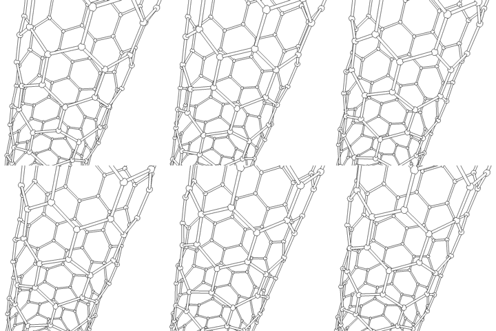
This animation of a rotating Carbon nanotube shows its 3D structure. Carbon nanotubes are among the numerous candidates for tissue engineering scaffolds since they are biocompatible, resistant to biodegradation and can be functionalized with biomolecules. However, the possibility of toxicity with non-biodegradable nano-materials is not fully understood.

To achieve the goal of tissue reconstruction, scaffolds must meet some specific requirements. A high porosity and an adequate pore size are necessary to facilitate cell seeding and diffusion throughout the whole structure of both cells and nutrients. Biodegradability is often an essential factor since scaffolds should preferably be absorbed by the surrounding tissues without the necessity of a surgical removal. The rate at which degradation occurs has to coincide as much as possible with the rate of tissue formation: this means that while cells are fabricating their own natural matrix structure around themselves, the scaffold is able to provide structural integrity within the body and eventually it will break down leaving the neotissue, newly formed tissue which will take over the mechanical load. Injectability is also important for clinical uses.

Many different materials (natural and synthetic, biodegradable and permanent) have been investigated. Most of these materials have been known in the medical field before the advent of tissue engineering as a research topic, being already employed as bioresorbable sutures. Examples of these materials are collagen or some linear aliphatic polyesters.

New biomaterials have been engineered to have ideal properties and functional customization: injectability, synthetic manufacture, biocompatibility, non-immunogenicity, transparency, nano-scale fibers, low concentration, resorption rates, etc. PuraMatrix, originating from the MIT labs of Zhang, Rich, Grodzinsky and Langer is one of these new biomimetic scaffold families which has now been commercialized and is impacting clinical tissue engineering.

A commonly used synthetic material is PLA - polylactic acid. This is a polyester which degrades within the human body to form lactic acid, a naturally occurring chemical which is easily removed from the body. Similar materials are polyglycolic acid (PGA) and polycaprolactone (PCL): their degradation mechanism is similar to that of PLA, but they exhibit respectively a faster and a slower rate of degradation compared to PLA.

Scaffolds may also be constructed from natural materials: in particular different derivatives of the extracellular matrix have been studied to evaluate their ability to support cell growth. Proteic materials, such as collagen or fibrin, and polysaccharidic materials, like chitosan or glycosaminoglycans (GAGs), have all proved suitable in terms of cell compatibility, but some issues with potential immunogenicity still remains. Among GAGs hyaluronic acid, possibly in combination with cross linking agents (e.g. glutaraldehyde, water soluble carbodiimide, etc...), is one of the possible choices as scaffold material. Functionalized groups of scaffolds may be useful in the delivery of small molecules (drugs) to specific tissues.


Types of Cells

Cells are often categorized by their source:

  • Autologous cells are obtained from the same individual to which they will be reimplanted. Autologous cells have the fewest problems with rejection and pathogen transmission, however in some cases might not be available. For example in genetic disease suitable autologous cells are not available. Also very ill or elderly persons, as well as patients suffering from severe burns, may not have sufficient quantities of autologous cells to establish useful cell lines. Moreover since this category of cells needs to be harvested from the patient, there are also some concerns related to the necessity of performing such surgical operations that might lead to donor site infection or chronic pain. Autologous cells also must be cultured from samples before they can be used: this takes time, so autologous solutions may not be very quick. Recently there has been a trend towards the use of mesenchymal stem cells from bone marrow and fat. These cells can differentiate into a variety of tissue types, including bone, cartilage, fat, and nerve. A large number of cells can be easily and quickly isolated from fat, thus opening the potential for large numbers of cells to be quickly and easily obtained. Several companies have been founded to capitalize on this technology, the most successful at this time being Cytori Therapeutics.
Mouse embryonic stem cells.  More lab photos
Mouse embryonic stem cells. More lab photos
  • Allogenic cells come from the body of a donor of the same species. While there are some ethical constraints to the use of human cells for in vitro studies, the employment of dermal fibroblasts from human foreskin has been demonstrated to be immunologically safe and thus a viable choice for tissue engineering of skin.
  • Xenogenic cells are those isolated from individuals of another species. In particular animal cells have been used quite extensively in experiments aimed at the construction of cardiovascular implants.
  • Syngeneic' or isogenic cells are isolated from genetically identical organisms, such as twins, clones, or highly inbred research animal models.
  • Primary cells are from an organism.
  • Secondary cells are from a cell bank.
  • Stem cells (see main article: stem cell) are undifferentiated cells with the ability to divide in culture and give rise to different forms of specialized cells. According to their source stem cells are divided into "adult" and "embryonic" stem cells, the first class being multipotent and the latter mostly pluripotent; some cells are totipotent, in the earliest stages of the embryo. While there is still a large ethical debate related with the use of embryonic stem cells, it is thought that stem cells may be useful for the repair of diseased or damaged tissues, or may be used to grow new organs.


Cells


Tissue engineering utilizes living cells as engineering materials. Examples include using living fibroblasts in skin replacement or repair, cartilage repaired with living chondrocytes, or other types of cells used in other ways.

Cells became available as engineering materials when scientists at Geron Corp. discovered how to extend telomeres in 1998, producing immortalized cell lines. Before this, laboratory cultures of healthy, noncancerous mammalian cells would only divide a fixed number of times, up to the Hayflick limit.

Extraction

From fluid tissues such as blood, cells are extracted by bulk methods, usually centrifugation or apheresis. From solid tissues, extraction is more difficult. Usually the tissue is minced, and then digested with the enzymes trypsin or collagenase to remove the extracellular matrix that holds the cells. After that, the cells are free floating, and extracted using centrifugation or apheresis.
Digestion with trypsin is very dependent on temperature. Higher temperatures digest the matrix faster, but create more damage. Collagenase is less temperature dependent, and damages fewer cells, but takes longer and is a more expensive reagent.


Tissue Engineering

Tissue engineering is the use of a combination of cells, engineering and materials methods, and suitable biochemical and physio-chemical factors to improve or replace biological functions. While most definitions of tissue engineering cover a broad range of applications, in practice the term is closely associated with applications that repair or replace portions of or whole tissues (i.e., bone, cartilage, blood vessels, bladder, etc...). Often, the tissues involved require certain mechanical and structural properties for proper function. The term has also been applied to efforts to perform specific biochemical functions using cells within an artificially-created support system (e.g. an artificial pancreas, or a bioartificial liver). The term regenerative medicine is often used synonymously with tissue engineering, although those involved in regenerative medicine place more emphasis on the use of stem cells to produce tissues.

In 2003, the NSF published a report entitled "The Emergence of Tissue Engineering as a Research Field" . which gives a thorough description of the history of this field.

Micromass cultures of C3H-10T1/2 cells at varied oxygen tensions stained with Alcian blue.

A commonly applied definition of tissue engineering, as stated by Langer and Vacanti, is "an interdisciplinary field that applies the principles of engineering and life sciences toward the development of biological substitutes that restore, maintain, or improve tissue function or a whole organ". Tissue engineering has also been defined as "understanding the principles of tissue growth, and applying this to produce functional replacement tissue for clinical use." A further description goes on to say that an "underlying supposition of tissue engineering is that the employment of natural biology of the system will allow for greater success in developing therapeutic strategies aimed at the replacement, repair, maintenance, and/or enhancement of tissue function.

Powerful recent developments in the multidisciplinary field of tissue engineering have yielded a novel set of tissue replacement parts and implementation strategies. Scientific advances in biomaterials, stem cells, growth and differentiation factors, and biomimetic environments have created unique opportunities to fabricate tissues in the laboratory from combinations of engineered extracellular matrices ("scaffolds"), cells, and biologically active molecules. Among the major challenges now facing tissue engineering is the need for more complex functionality, as well as both functional and biomechanical stability in laboratory-grown tissues destined for transplantation. The continued success of tissue engineering, and the eventual development of true human replacement parts, will grow from the convergence of engineering and basic research advances in tissue, matrix, growth factor, stem cell, and developmental biology, as well as materials science and bioinformatics.